A cell-mapping scientist’s two-continent path
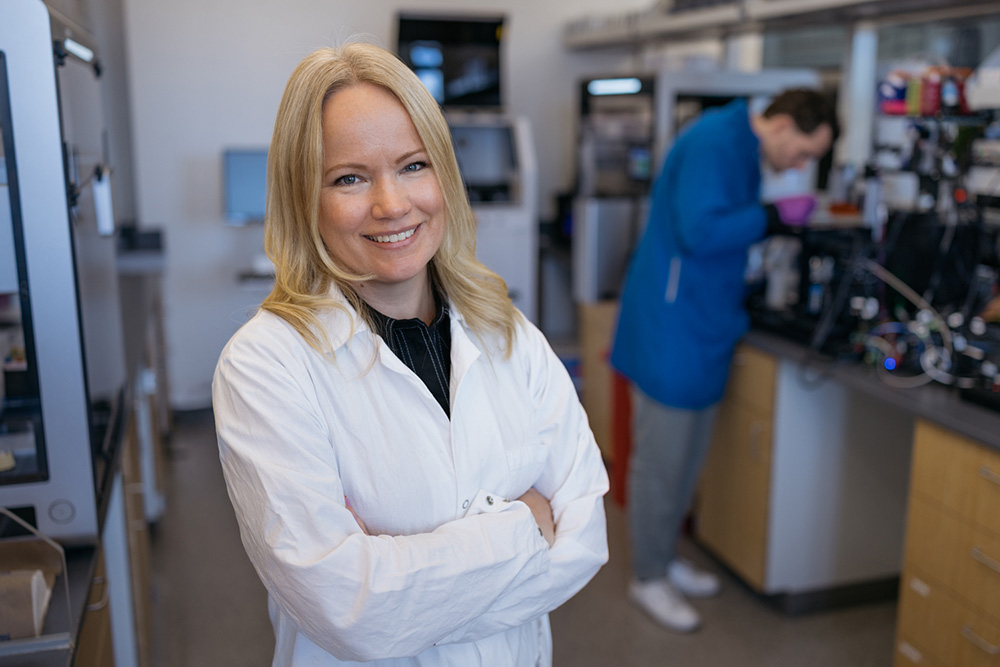
From Stanford’s sunlit engineering quad to SciLifeLab’s brisk knowledge hub in Stockholm, Emma Lundberg works across two distinct innovation cultures. She sees lessons for Sweden in Silicon Valley’s close links between academia, investors, and industry, but also strengths at home that should not be lost.
September 14, 2026


Beyond Stanford’s historic sandstone arcades, four modern buildings frame the central courtyard of the Science and Engineering Quad. The largest, the Shriram Center for Bioengineering & Chemical Engineering, covers more than 19,000 square meters and contains 34 specialized laboratories, comparable in scale to SciLifeLab Campus Solna, which spans approximately 22,500 square meters. Scattered across the courtyard are 12 enormous polished stone spheres. They form Pars pro Toto, an installation by the Polish-German artist Alicja Kwade, created from stones sourced from different parts of the world.
As we crossed the courtyard, Emma Lundberg pointed out one of the spheres. It was made from Swedish granite – a small piece of Sweden’s geological history resting beneath the California sun. She showed me around the laboratory and introduced me to several members of her team. Here, she says, academia, technology companies, investors, and non-profit organizations overlap in ways that help ideas move easily.
Stanford vs. Stockholm
Emma Lundberg is Professor at KTH Royal Institute of Technology and Associate Professor of Bioengineering and of Pathology at Stanford University. She also plays a key part in The Human Protein Atlas, where she leads the subcellular profiling work. When I ask her what the biggest difference is between the two environments, she points out, for example, that in Stanford there is an ecosystem of academia, non-profits, and industry in the life sciences and tech bio space that works very well.
Things move quickly, investments and bets are large, and there is a Silicon Valley mindset of dreaming big. That shapes how you think about risk, for both good and bad. Different things may grow better at different paces, and it is interesting to learn how to operate in both.
“I would love to see more of that in the Stockholm region,” she says. “Certain things are done better in academia and certain things are done better in industry. There is nothing wrong with professors spinning out companies if translation into clinical care is better done there. That mindset is more established here.”
This is also a very particular, slightly crazy part of the world – the universe spins faster, she adds. “Things move quickly, investments and bets are large, and there is a Silicon Valley mindset of dreaming big. That shapes how you think about risk, for both good and bad. Different things may grow better at different paces, and it is interesting to learn how to operate in both.”
I also ask her if the formal support for commercialization is better at Stanford, or if the difference is mainly cultural.
“The intellectual property systems are different. In Sweden, the teacher’s exemption means researchers generally own their innovations. That is a great freedom, but it can also mean that finding the funding to protect an invention relies on yourself. At Stanford, the university owns inventions made under its policies, but it also takes on patenting and provides strong support. The researcher’s ownership is smaller, but the process can move faster,” she says.
The biggest difference, however, is probably the network, she adds. “Everyone knows someone in venture capital or someone who has started a company. Venture capitalists monitor universities, and many bioengineering students arrive with a dream of building a company. In Sweden, the university and start-up environments are more separated.”
Mapping the cell as a system
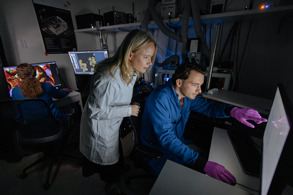
Lundberg’s laboratory remains rooted in basic biology, combining assay development, computational modeling, and biological application. She describes the team as working at the intersection of AI, computer vision, microscopy, and spatial biology.
“We want to understand how the cell functions as a system. A cell has around 20,000 proteins, and proteins are the executors of function. If we understand how they work together, which proteins interact and how they form networks, then we can begin to model cellular function. Our niche is spatial and subcellular biology. Cells are not bags of molecules. They are highly organized, with different organelles where specialized functions take place. We need to understand that three-dimensional structure, and how it reorganizes over time, if we really want to understand the cell,” she says.
So how can such fundamental mapping become useful to drug discovery?
“Most of the data we generate is published in the Human Protein Atlas. We know that all the major pharma companies and many biotech startups use it. That is why I strongly believe in open science. The data may initially reveal basic biological insights, but it can be mined through many different hypotheses. By sharing it openly, we can accelerate drug discovery indirectly,” says Lundberg.
Her collaborations also move more directly toward application. A Danaher Beacon research collaboration has the aim of improving cancer drug screening. Lundberg describes the broader ambition as developing four-dimensional phenotyping of organoids, capturing biological structure and treatment response over time. A recent preprint describes another key project, ProtiCelli. Trained on 1.23 million Human Protein Atlas images, the generative model simulates microscopy images for approximately 12,800 human proteins using only three cellular landmark stains.
Virtual cells, without the hype
Virtual-cell models are attracting major scientific and commercial attention, and Lundberg welcomes the momentum but distinguishes potentially useful models from the more distant ambition of creating a complete digital replica of human biology.
What has changed now, and where do you expect the first real impact?
“The dream of building models of cells is old. What has changed is the tools. We can generate biological data at a very large scale, and AI models can learn patterns from it,” she says. “But I think we are still far from a truly high-fidelity biological simulator. Biology spans many spatial and temporal scales, and we lack data across many modalities. The first impact will not come from replacing the wet lab. What excites me is a lab-in-the-loop system.Today, we often run a very large and expensive experiment and analyze it afterwards. In the future, we may run a smaller set, use models to simulate possibilities and choose the most informative next experiments, then update the model and iterate. The models do not have to be perfect to be useful,” she says.
The dream of building models of cells is old. What has changed is the tools. We can generate biological data at a very large scale, and AI models can learn patterns from it.
Lundberg sees SciLifeLab’s Alpha Cell program and parallel international initiatives by BioHub and others as complementary rather than as a race with a single winner. There are too many cell types, disease states, and biological scales for one program to cover alone. Her greater concern is fragmentation: large datasets being produced in different parts of the world without sufficient coordination to allow future models to learn from them together.
Trust begins with the antibody
However, before an image can train an AI model, the measurement behind it must be trustworthy.
“An antibody is a proxy measurement. If you have an antibody for protein X, you must be very sure that it actually binds protein X. Otherwise, you may build hypotheses on false or misleading data,” says Lundberg. “This is something we have always pushed hard in the Human Protein Atlas. It is not only that the antibodies are highly validated; we also publish the validation data so users can make their own assessment. And when users challenge a result, we listen. That feedback is an important part of maintaining quality.”
A researcher may find a protein in the Atlas that is particularly interesting and wants to study it in a rare-disease cohort. Antibodies developed through the project are available through Atlas Antibodies.
“If the antibody were not available, they might need to generate a new one or obtain it directly from an academic laboratory, which cannot work at scale. Making the antibodies available allows researchers to use the Human Protein Atlas not only as a knowledge base, but as a starting point for their own work,” says Lundberg.
Making knowledge usable
The landmark Subcellular Atlas, published in Science in 2017, illustrates the value of this foundation. It mapped the locations of more than 12,000 proteins across organelles and other structures inside human cells.
Approximately half of the proteins were found in more than one cellular compartment, supporting the idea that many proteins may perform different functions depending on where they are located. For thousands of proteins, the project also provided the first evidence of where they could be found inside human cells. The result was a reference map that researchers around the world could use to generate new questions.
The universe may spin faster in California. But a piece of Sweden is already part of its orbit.
Yet a map has greater impact when other scientists can build on it. Open data allows a researcher to identify a potentially important protein. Access to the underlying tools allows that researcher to investigate it in another cell type, disease, or patient group.
Walking back into the courtyard, the stone spheres offered a fitting final image. The title Pars pro Toto – “a part for the whole” – could also describe the kind of research ecosystem Lundberg is advocating. Each participant brings a different history, capability, and perspective, but their value increases when they become part of something larger. The universe may spin faster in California. But a piece of Sweden is already part of its orbit.
Published: September 14, 2026